Book an Appointment
CECT For Axilla
Medifyhome has collaborated with the best pathology laboratories that are NABL and NABH certified and follow ISO safety guidelines to provide the best CECT For Axilla at an affordable price for needy individuals. Contrast-enhanced computed Tomography (CECT) of the Axilla is a strong imaging modality that assesses the axillary region, or armpit, which is very important in diagnosing many conditions, especially those involving lymph nodes and soft tissues. The axilla contains vital lymphatic structures that may be involved in diseases such as breast cancer, lymphoma, infections, and other types of tumors. CECT combines the detailed imaging power of a standard CT scan with the enhanced clarity provided by a contrast dye, allowing healthcare professionals to obtain high-resolution images of both the soft tissue and the bone structures in this complex area. CECT is especially useful in the evaluation of axillary lymph nodes, which can become enlarged or abnormal due to metastatic spread, infections, or other pathologies. Whether it is used to detect the spread of breast cancer to lymph nodes, identify the presence of an abscess, or evaluate other soft tissue masses, CECT provides invaluable insights that guide diagnosis, treatment planning, and follow-up care. This non-invasive imaging method is vital in staging cancers, monitoring the effectiveness of treatments, and detecting any recurrence of disease.
To schedule an appointment for a CECT for the Axilla, simply contact Medifyhome or call our customer care at +919100907036 or +919100907622 for more details and queries.
What is CECT?
CECT is an acronym referring to Contrast-Enhanced Computed Tomography, a form of medical imaging technique to get clear images within one’s body to diagnose or keep track of a variety of health conditions. In the use of CECT, an injecting contrast agent, usually via a vein, serves as a contrast dye that contrasts the blood vessels, tissue, and organs that take place in a CT scan. The contrast material provides contrast enhancement of structures in the scan, such that abnormalities can be more easily identified, whether it’s tumors, infection, or vascular diseases. CECT is normally used in identifying and monitoring cancers, investigating blood vessel disease (an aneurysm, for example), investigating any internal injuries resulting from a trauma, or the monitoring of infections/inflammatory diseases, among others. The use of a contrast gives enhanced clarity with details, thus aiding in proper medical diagnostic tools. A standard CT scan uses X-rays to create cross-sectional images, known as slices, of the body. The images are then put together to form a 3D image that can help doctors see internal organs and structures. CT scans are usually quick, and when contrast is added, results are obtained in a relatively short time.
Role of CECT for Imaging the Axilla
Contrast-enhanced computed Tomography (CECT) is a potent imaging modality applied for axillary assessment, the area below the shoulder joint which holds various important structures, including lymph nodes, blood vessels, nerves, and adipose tissue. The key roles and benefits of using CECT in axillary imaging are described below:
- Evaluation of Lymph Node Involvement:
- Axillary Lymphadenopathy: The axilla is one of the most common sites for lymph nodes that are involved in cases like breast cancer, lymphoma, infections, and sarcoidosis. CECT provides for the assessment of lymphadenopathy, giving information regarding lymph node size, shape, and density.
- Characterization of the Lymph Node: With CECT, malignant vs. benign lymph nodes could be differentiated by evaluation based on morphology, enhancement, and central necrosis.
- Tumor Detection and Staging :
- Primary Tumor Visualisation: CECT is particularly helpful in the staging of breast cancer, as it can show tumor invasion into the axillary lymph nodes or adjacent tissues. It provides detailed views of the breast tissue, axillary nodes, and nearby structures, enabling the radiologist to assess the extent of the tumor.
- Tumor Invasion: In locally advanced breast cancer or sarcoma, CECT may reveal the extent of invasion by the tumor into the surrounding tissue, including axillary fat, muscles, or blood vessels. This helps in planning surgical resection or other treatments.
- Vascular Assessment:
- Blood Vessels in the Axilla: The axilla contains important vascular structures, including the subclavian artery, axillary artery, and associated veins. CECT can provide detailed images of these blood vessels, helping to assess conditions like vascular malformations, aneurysms, or thrombosis.
- Tumor Vascularity: In cases of axillary region tumors, it can evaluate the vascularity (blood supply) of the tumor. Usually, malignant tumors have a richer vascularity compared with a benign mass. In a situation like this, a differentiation between benign and malignant could be made using this vascularity.
- Determination of Infections and Abscesses
- Axilla infections An infection may be caused by either the bacteria or the fungi. Abscess, cellulitis, etc., in this region can be visualized with the help of CECT, which gives an idea as to the extent of infiltration and severity of infection.
- Abscess Drainage: In enlarged axillary abscesses, CECT can be useful to guide drainage procedures by showing the accurate location and size of the abscess to help surgeons or radiologists during needle aspiration or drain placement.
- Axillary Mass Evaluation
- It includes benign masses as well as malignant tumors. CECT has to be done in evaluating any abnormal mass in the axillary region, which provides characterisation of the mass concerning its size, enhancement pattern, and relation to other surrounding structures.
Procedure for CECT of the Axilla
- The process of Contrast-Enhanced Computed Tomography (CECT) of the axilla is performed by following a series of steps to obtain high-quality images of the axillary region, which includes the axillary lymph nodes, vessels, muscles, and soft tissues around them. Below is an outline of the typical procedure for performing a CECT of the axilla:
- Pre-Procedure Preparation:
- Before the procedure, certain steps are followed to ensure the patient’s safety and optimize the imaging quality.
- Medical History: The patient will be required to provide a comprehensive medical history, which includes the following:
- All allergies, especially contrast agents.
- The kidney function because contrast agents affect the renal function.
- If pregnant or breastfeeding, they have to inform the doctor as CT scans include ionizing radiation, and such radiations are not usually done during pregnancy except if necessary.
- Fasting: Generally, fasting is not necessary before a CECT of the axilla, except when the scan is done in conjunction with imaging of the chest or abdomen. In such cases, fasting for about 4–6 hours may be recommended to minimize the risk of nausea when the contrast is administered.
- Hydration: Some patients will be instructed to drink significant amounts of fluids before the test, especially if they suffer from kidney disease, to wash out the contrast agent after scanning.
- Clothing and Metals: The patient will be required to change into a hospital gown and remove jewelry, watches, or any other metal objects. Any metal can interfere with CT imaging.
- Positioning the Patient:
- The patient will be put on the CT scan table. For a CECT of the axilla, the position for the patient is lying in a supine position most of the time, except for some clinical indications requiring scanning of other parts; the patient may extend arms above their head or on the sides to allow direct exposure of the axilla.
- In some cases, arm positions may differ depending on the field of view and whether both axillae are being simultaneously imaged.
- CT Scanning:
- Scanning Beginning: Once the contrast agent has been administered, scanning will begin. The large circular machine of the CT scanner includes an X-ray tube that rotates around the body while the patient passes through the opening of the scanner (the gantry).
- Timing: The timing of contrast injection is critical to catch the proper enhancement of tissues. For some scans, it requires rapid infusion of contrast to capture the vascular structures. For example, to detect tumors or lymph nodes. For others, a slower infusion might be needed.
- Scan Time: The actual scan time is usually around 5-15 minutes, depending upon the area to be covered and whether multi-phase scanning is required (for instance, arterial and venous phases).
- The table goes through the CT scanner.
- The patient is to lie as still as possible for the scan, though they may need to hold their breath in a few spots to reduce motion artefacts and get clear images.
Benefits of CECT for Axillary Imaging
Contrast-enhanced computed Tomography (CECT) axillary imaging holds a significant place in diagnosing, staging, and managing several conditions involving the axillary region, which include the axillary lymph nodes, vessels, muscles, and the surrounding soft tissues. Key benefits of CECT axillary imaging include the following:
- Enhanced Visualization of Lymph Nodes:
- Detection of Lymphadenopathy One of the key uses of CECT in axillary imaging is the detection of lymph node enlargement (lymphadenopathy), which is suspected to be related to infection, lymphoma, or metastatic disease from primary malignancies, including breast cancer. Lymph nodes can then easily become visualized due to the enhanced contrast agent for any pathology related to malignancy (eg, malignant enlargement) and necrosis.
- Differentiation of Benign and Malignant Nodes: CECT helps differentiate benign from malignant lymphadenopathy by assessing the morphology, enhancement patterns, and presence of central necrosis.
- Assessment of Tumors and Masses:
- Detection of Primary and Secondary Tumors: CECT is useful in identifying primary tumors in the axilla, such as soft tissue sarcomas or neurogenic tumors, and secondary tumors (metastasis) that may spread from adjacent structures (such as the breast).
- Tumor Vascularity: The contrast agent has the advantage of better opacification of blood vessels, which is more helpful to assess vascularity in a tumor.
- Detection of Infection and Abscesses:
- Axillary Infections: CECT can be very useful for infections occurring in the axillary regions, such as abscesses, cellulitis, or infected lymph nodes. Infected areas show high contrast enhancement and may present with fluid collections (pus) that can be localized.
- Guiding drainage procedures: CECT guides percutaneous drainage procedures in large or complex abscesses. It allows the interventional radiologist or surgeon to plan the procedure by showing the size, location of the abscess, and surrounding structures.
- Preoperative planning for axillary surgery
- Surgical Planning for Breast Cancer: For patients with breast cancer or other malignancies involving the axilla, CECT is used to plan axillary lymph node dissection (ALND) or other surgical interventions. By clearly defining the location, size, and number of involved nodes, CECT can help in deciding the extent of surgery and avoid unnecessary removal of healthy tissue.
- Evaluation of Vascular Abnormalities:
- Blood Vessel Visualization: There are several key blood vessels in the axilla, including the axillary artery, subclavian artery, and brachial artery. CECT improves the visualization of these structures, making it easier to identify abnormalities such as:
- Arteriovenous Malformations (AVMs): Abnormal connections between arteries and veins that may lead to bleeding or other complications.
- Vascular Compression or Thrombosis: CECT helps identify the blockage (thrombosis) or compression of blood vessels affecting the free flow of blood towards the arm or breast.
- Test Type: CECT For Axilla
- Preparation:
- Wear a loose-fitting cloth
- Minimum 4 hours fasting required
- Carry Your ID Proof
- Prescription is mandatory for patients with a doctor’s sign, stamp, with DMC/HMC number; as per PC-PNDT Act
- Reports Time: With in 3-4 hours
- Test Price: Rs.2500
How can I book an appointment for a CECT for the Axilla through Medifyhome?
To schedule an appointment for a CECT for Axilla, simply contact Medifyhome or call our customer care at +919100907036 or +919100907622 for more details and queries.
What is CECT, and how is it different from a standard CT scan?
CE is referred to as CECT. In other words, a type of contrast-enhanced computed tomography utilizes a contrast dye in the diagnosis to enhance the appearance of blood vessels, tissues, and abnormalities. Contrary to the regular CT scan that offers detailed images of the bones and organs, the clear and precise images provided by CECT render it more valuable in diagnosing and assessing areas like the axilla, where assessment of lymph nodes and soft tissue masses predominates.
Why is CECT employed for imaging the axilla?
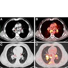
CECT is applied to investigate diseases involving the axilla, including enlarged lymph nodes (infections, metastasis, or lymphoma), soft tissue tumors, and abscesses. It is of particular importance in breast cancer, as the status of axillary lymph nodes is important for the determination of the spread of disease and treatment modalities.
Is the procedure of CECT painful?
No, the actual CECT scan is not painful. A CT scanner takes detailed images as the patient lies on a table. The contrast dye is injected into a vein. The patients may feel some warmth in their veins or a metallic taste in the mouth, which subsides after some time and isn’t painful at all.
What are the risks for CECT for the axilla?
While CECT is a relatively safe procedure, there are some risks. The contrast dye used in the procedure can cause allergic reactions in some individuals, ranging from mild symptoms like itching or nausea to more severe reactions. Patients with kidney disease may need to avoid CECT as the contrast agent can strain kidney function. So, it is essential to inform your doctor if you have any allergies or pre-existing medical conditions before undergoing the scan.
How long does a CECT scan of the axilla take?
The actual CECT scan typically takes about 10 to 15 minutes. However, the entire process, including preparation (such as administering the contrast dye) and post-procedure care, may take around 30 minutes.
Are there any special preparations required before a CECT scan?
You may be asked to fast for a few hours before the procedure, especially if you are going to receive an intravenous contrast agent. You should inform your doctor about any allergies, especially contrast dyes or iodine, and any medical conditions, such as kidney problems or pregnancy. You should also avoid wearing any metal items, such as jewelry, in the area being scanned.
Can CECT detect axillary cancers?
Yes, the CECT can detect changes that are associated with cancer in the axilla. In other words, it is used for breast cancer that has extended to axillary lymph nodes. This modality provides information regarding size, shape, and the extent of tumor or enlarged lymph nodes which helps to stage the tumor and make plans for its treatment.
Most Booked Ct Scan Parts
With Medifyhome, book a CT scan get the fastest appointment scheduling at NABH & ICMR Certified diagnostic centers near you. Trusted reports, expert radiologists, and seamless service-only a click away!
Why Choose Medifyhome for CECT For Axilla?
Medifyhome is an online medical consultant that provides home-based medical services not only in your area but also in most cities in India, including Hyderabad, Chennai, Mumbai, Kolkata, and more. We have collaborated with diagnostic centers that have the best machines and equipment to ensure you get accurate results. Medifyhome provides 24-hour customer service for booking the appointment of the services and guides you with instructions. Medifyhome also provides the best diagnostic centers at low prices. Once you receive your test results, you can easily book an appointment with our network of experienced doctors for consultation. To schedule an appointment for a CECT for Axilla, simply contact Medifyhome or call our customer care at +919100907036 or +919100907622 for more details and queries.


