
MRI For Cardiac
Book your MRI For Cardiac at Medifyhome, where we offer top-quality scans at the best prices. Our NABL & NABH accredited diagnostic centers ensure accurate and reliable results.


Book an Appointment
MRI For Cardiac
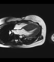
Medifyhome has collaborated with the best pathology laboratories that are NABL and NABH certified and follow ISO safety guidelines to provide the best MRI For Cardiac at an affordable price for needy individuals. Cardiac MRI is one of the leading-edge, non-invasive imaging technologies capable of providing crucial diagnosis for heart conditions. Unlike the routine X-rays or CT scanning, the use of strong magnet and radio waves by MRI produces clear and high-resolution imaging of the heart’s morphology, its function, or blood flow; in other words, it helps to visualize quite a broad range of possible cardiovascular diseases.
Whether it is about heart disease, congenital heart defects, or any other heart concern, a cardiac MRI can provide a wide view of the heart’s status, and this helps doctors get accurate diagnoses and create effective treatment plans. Cardiac MRI does not involve radiation; it has the ability to detect the slightest changes within the tissue of the heart, so it is the safest and most effective method for diagnosis and management of heart conditions. To schedule an appointment for a MRI For Cardiac, simply contact Medifyhome or call our customer care at +919100907036 or +919100907622 for more details and queries.
Overview
Specification
FAQ
Overview
How Does Cardiac MRI Work?
Cardiac MRI, or Magnetic Resonance Imaging, is a non-invasive technique in imaging. It uses powerful magnetic fields, radio waves, and computer processing to create detailed images of the heart and surrounding blood vessels. Here’s how it works, step by step:
- Creation of Magnetic Field:
The MRI machine generates a strong magnetic field, usually in the range of 1.5 to 3 Tesla, which is 30,000 to 60,000 times stronger than Earth’s magnetic field.
This magnetic field aligns the protons (hydrogen nuclei) in the body, which are abundant in water and tissue, including the heart muscle.
- Radiofrequency Pulses:
Once the protons in the body are aligned, the MRI machine sends short bursts of radiofrequency (RF) pulses. These RF pulses “flip” the protons out of alignment.
When the RF pulses are switched off, the protons begin to relax back in their alignment, which then releases energy.
- Signal Detection:
The signal is picked up by the MRI machine’s receiver coils where it converts into data.
Other tissues within the body that include muscles, fats, or blood emit varying levels of energy while returning to alignments, hence helping in identification of the various tissues within the heart.
- Reconstruction of Images:
The signals are processed by a computer to produce detailed cross-sectional images or “slices” of the heart.
These images can be displayed in several planes (horizontal, vertical, and oblique) and provide information on the structure and function of the heart.
- Cardiac Specific Imaging:
Cardiac MRI may also include various imaging sequences to assess specific components of heart health:
Cine MRI: It is an imaging type that captures the heart in motion, thus allowing assessment of heart wall motion, function, and volume changes in the cardiac cycle (systole and diastole).
Late Gadolinium Enhancement (LGE): A contrast agent-gadolinium is infused into the bloodstream, thus highlighting areas of the heart with scar tissue or damage such as after a heart attack.
What Conditions Can Cardiac MRI Diagnose?
Cardiac MRI is a very versatile imaging technique that can diagnose the vast majority of cardiovascular diseases. It gives detailed information about the structure and function of the heart, making it possible to diagnose congenital and acquired heart diseases. Some of the most common conditions diagnosed by cardiac MRI include the following:
- Coronary Artery Disease (CAD) and Myocardial Infarction (Heart Attack)
Scar Tissue: After a heart attack, MRI of the heart with late gadolinium enhancement might reveal areas of scar tissue within the heart muscle, suggesting myocardial infarction.
Viability evaluation: Using MRI, it is also possible to evaluate the viability of cardiac tissues in which a damaged region of heart muscle either remains viable and functional or permanently and irreversibly damaged.
- Cardiomyopathy
Dilated Cardiomyopathy (DCM): MRI is helpful in the assessment of cardiac enlargement and estimation of the function of the pumping ability of the heart, i.e., ejection fraction. It also detects the areas of fibrosis and structural abnormalities of the heart muscle.
Hypertrophic Cardiomyopathy (HCM): MRI measures the thickness of the heart muscle, particularly of the left ventricle, and identifies asymmetrical thickening of the wall of the heart, a hallmark of HCM.
Restrictive Cardiomyopathy: MRI can assess the stiffness and thickening of the heart muscle that impedes proper blood flow and reduces heart function.
- Congenital Heart Disease
Structural Abnormalities: MRI provides detailed images of congenital heart defects, such as septal defects (holes between the heart’s chambers), valve abnormalities, and malformations of the heart’s structure. It is especially useful for assessing complex congenital conditions.
Evaluation of Atrial Septal Defect (ASD) and Ventricular Septal Defect (VSD): MRI can measure the size of defects and any resultant disturbance in blood flow.
- Heart Valve Disease
Valve Insufficiency: MRI can determine the size of heart valves affected with stenosis (reduction in size) or regurgitation (leakage). MRI can also provide information about the impact of valve dysfunction on heart function.
Valvular Calcification: MRI can assist in identifying calcifications or other alterations of the heart valves, which might lead to stenosis.
- Pericardial Disease
Pericarditis: MRI can be utilized in assessing inflammation of the pericardium by determining signs of inflammation or thickening of the pericardium.
Pericardial Effusion: MRI can detect fluid accumulation around the heart due to pericarditis, trauma, or other conditions.
Constrictive Pericarditis: MRI may be able to detect abnormal thickening or calcification of the pericardium that limits cardiac function.
Benefits of Cardiac MRI
Cardiac MRI offers several unique advantages over other imaging modalities for the assessment of heart diseases. Here are the benefits that come with cardiac MRI:
- High-resolution detailed imaging
Superior image quality: Cardiac MRI produces images with high resolution and detail about the heart’s structure, which makes the muscle of the heart, valves, blood vessels, and surrounding tissues well visualized.
Multiplanar Imaging: Unlike other imaging modalities, MRI can create images in any plane, offering a panoramic view of the heart.
Tissue Characterization: MRI can distinguish between several types of tissue, such as normal myocardium (heart muscle), fat, scar tissue, and edema (swelling), using advanced imaging techniques like Late Gadolinium Enhancement (LGE) or T1/T2 mapping.
- Non-Invasive and No Radiation
No Radiation Exposure: CT scans or X-rays involve ionizing radiation. MRI does not, hence a safer method for repeated evaluation, especially in younger patients or those requiring long-term follow-up.
Non-Invasive: It does not require any invasive procedures like catheterization or biopsies to assess cardiac conditions.
- Thorough Heart Function and Structure Evaluation
Functional Imaging: Cardiac MRI provides accurate measurement of ejection fraction, cardiac output, and ventricular volumes, all of which are necessary for the assessment of heart function, especially in diseases such as heart failure.
Cine MRI: It allows imaging of the motion of the heart, thereby allowing the evaluation of dynamic heart function at any point during the cardiac cycle (both systole and diastole) and heart wall motion and regional dysfunction.
- Identification of Scar Tissue and Myocardial Viability
Myocardial Infarction (Heart Attack): MRI with Late Gadolinium Enhancement (LGE) is the gold standard for detecting myocardial infarction and identifying areas of myocardial scarring (fibrosis) that may result from a heart attack.
Assessment of Viability: MRI can assess the viability of heart muscle after myocardial infarction or other forms of myocardial damage for appropriate treatment, like whether a patient needs revascularization.
- Very useful to identify inflammation of the myocardium
Myocarditis: Cardiac MRI remains one of the best imaging modalities for visualizing myocarditis. It includes detection of inflammation, swelling, and fibrosis involved in diagnosing and monitoring the progressive course of this disease.
Sarcoidosis and Other Inflammatory Conditions: MRI is helpful in delineating cardiac sarcoidosis when clusters of inflammatory cells infiltrate the myocardium, which may lead to arrhythmias or heart failure.
Preparation for Cardiac MRI
Preparing for a Cardiac MRI is usually straightforward, but there are a few key steps that will help ensure the procedure goes smoothly. Here’s what you can expect and how to prepare for the exam:
- Pre-Screening and Health History
Health Information: Before the MRI, you will be asked to provide detailed health information, including any medical conditions, medications you are taking, and any history of allergies.
Metal Implants: Because MRI machines operate on strong magnetic fields, you should let the technician or your doctor know if you have any metal implants or devices, such as:
- Pacemakers or defibrillators
- Artificial heart valves
- Stents
- Joint replacements, screws, or plates
- Clips or coils used in brain surgery or other treatments
- Tattoos with metallic ink or piercings
2. Pregnancy: Although MRI is considered safe, it is usually avoided in the first trimester unless absolutely necessary. So, do not forget to inform the healthcare provider whether you are pregnant or a breastfeeding mother.
Food and Drink Instructions
Fasting: If your Cardiac MRI will involve a contrast agent, you may be asked to fast for 4 to 6 hours before the test. This is so that your stomach is empty, and the risk of nausea or other side effects is minimized.
Normal Meals: In most cases, fasting is not necessary, but it’s always a good idea to confirm with your healthcare provider or the MRI facility for specific instructions.
- Clothing and other personal items
Loosely fitting, comfortable, non-metallic clothing No metal fasteners Loose shirts, skirts, and open-toe shoes You are asked to change into the hospital gown to remove metallic items that may interfere with the MRI machine.
Take away Jewelry and Personal Effects: Remove all jewelry, wristwatch, piercings, hairpins, and other metal objects prior to undergoing a test. These might disturb the MRI images or cause harm in the magnetic field.
- Medication
Continue Routine Medications: Most patients do not need to stop routine medications before the MRI. If you are on heart medications, such as blood pressure or heart failure medications, you can usually take them as prescribed unless your doctor has told you otherwise.
- What to Expect During the MRI
Duration: The procedure usually takes 30 to 90 minutes, depending on the complexity of the exam. Cardiac MRIs tend to take longer than standard MRIs because they often involve both imaging the heart’s structure and function, as well as using contrast.
Lie flat on your back on the MRI table, which then moves into the scanner. Try to remain motionless throughout the procedure. It may be necessary for you to be given instructions to help control your breathing as a means of minimizing movement. In some cases, some breath holding may be necessary in order to get a good view of the heart when the images are being obtained.
Noise: MRI devices are noisy. You can expect to receive earplugs or headphones during the MRI to dampen the sounds. In addition, the radiology technician may supply you with music to help distract you from the scanner.
Specification
- Test Type: MRI For Cardiac
- Preparation:
- Wear a loose-fitting cloth
- You will be asked to fast for 4 hours prior to your exam.
- Carry Your ID Proof
- Prescription is mandatory for patients with a doctor’s sign, stamp, with DMC/HMC number; as per PC-PNDT Act
- Reports Time: With in 4-6 hours
- Test Price: Rs.8000
FAQ
How can I book an appointment for a MRI For Cardiac through Medifyhome?
To schedule an appointment for a MRI For Cardiac, simply contact Medifyhome or call our customer care at +919100907036 or +919100907622 for more details and queries.
Is Cardiac MRI Safe?
Yes, Cardiac MRI is generally considered very safe. It uses magnetic fields and radio waves, which do not involve radiation, unlike other imaging techniques such as X-rays or CT scans. However, certain patients with metal implants, such as pacemakers or defibrillators, may not be eligible for an MRI. It’s important to inform your healthcare provider about any implants or devices before the procedure.
Does Cardiac MRI Hurt?
No, a Cardiac MRI is a painless procedure. During the scanning, you will have to lie still for some time when the images are taken. The machine can produce very loud noises, but you will be provided ear protection to reduce the sound. If a contrast agent is used, you might experience a mild, temporary sensation of warmth.
How Long Does a Cardiac MRI Take?
The length of a typical Cardiac MRI scan is between 30 to 60 minutes. If more complex images are needed, the scan might take longer. Some may even take longer if they need multiple views or require some advanced techniques.
Can I Eat or Drink Before My Cardiac MRI?
You will usually be told to refrain from eating and drinking for several hours prior to your Cardiac MRI. This is often the case if you are going to be given a contrast dye, but this again depends on the procedure being carried out. Be sure to observe the specific requirements of your doctor or the imaging center.
Is There Any Danger of Contrast Dye During Cardiac MRI?
Overall, the contrast dye used during a Cardiac MRI is relatively safe. However, in some patients, there is a mild side effect associated with it, such as a metallic taste in the mouth, warmth, and a slight headache. Individuals with kidney problems or having a known allergy to agents used for contrast should disclose it to the doctor before scheduling the scan so that there can be an alternative solution and precautions can be taken.
How Should I Prepare for a Cardiac MRI?
Preparation for a cardiac MRI is generally straightforward. You will be asked to remove your clothes and wear a hospital gown and remove all metallic objects such as jewelry, watches, or hairpins. If a contrast agent is to be administered, you will be requested to abstain from food and fluids for several hours before the test. They may also inquire about any current medical conditions, allergies, or implants.
Will I Need to Follow Up After My Cardiac MRI?
After your Cardiac MRI, your doctor will review the results and share the findings with you. Depending on the results, follow-up appointments may be necessary to discuss treatment options, further testing, or monitoring your heart condition over time.
How Soon Do I Get My Results?
The results of the MRI will be available in a matter of days after the examination. In case the urgency requires it, your physician will receive preliminary findings before this time. Your doctor will contact you to discuss the results and next steps.
Why Choose Medifyhome for MRI For Cardiac?
Medifyhome is an online medical consultant that provides home-based medical services not only in your area but also in most cities in India, including Hyderabad, Chennai, Mumbai, Kolkata, and more. We have collaborated with diagnostic centers that have the best machines and equipment to ensure you get accurate results. Medifyhome provides 24-hour customer service for booking the appointment of the services and guides you with instructions. Medifyhome also provides the best diagnostic centers at low prices. Once you receive your test results, you can easily book an appointment with our network of experienced doctors for consultation. To schedule an appointment for a MRI For Cardiac, simply contact Medifyhome or call our customer care at +919100907036 or +919100907622 for more details and queries.