How Does 3D CT Orbit Work?
A 3D CT Orbit is the advanced scan, combining scanning technology using CT with improved reconstruction methods to help obtain high resolution and detailed 3-D images of the orbital area. Let’s rephrase it to:
- CT scan or cross-sectional image process
X-ray Technology: This utilizes the application of a CT scanner, which owns various cross-sectional (2D) images or slices of the orbital region, using X-ray technology. The scanner will thus be able to capture those different angles and, hence, image the inner structures of the orbit in detail.
Rotating X-ray tube: In the scanning system, the patient does not move, and a rotating X-ray tube emits x-rays that hit inside an array of sensors or detectors attached to the side of the scanner.
Data Acquisition: Each X-ray beam passing through the body is absorbed at different rates by the different tissues in the orbit. The harder structures like bones absorb more X-rays than the softer tissues that include muscles and blood vessels. The detectors measure the X-rays that pass through and transmit this information to a computer for processing.
- 3D Reconstruction
Image formation The computer reconstructs the information it receives from the scanner into a 2D cross-sectional image of the orbital area. In this sense, this is indeed a cross-section of the given area, similar to cutting a bread roll into pieces.
Three-dimensional rendering. With all these cross-sections in 2D, all this information is compiled, and then it is assembled with computer software to take on a complete model in 3D. This gives much more extended, multi-dimensional views of an eye socket, muscles, nerves, and all the surrounding structures.
Viewing from Multiple Perspectives: Once the 3D image has been created, the radiologist or physician may also rotate the model for enhanced examination and zoom in on interesting features. This provides a magnificently detailed and clear view of the anatomy within the orbit.
- Patient Positioning
Patient Position: The patient will lie on the table. As scanning is done, the head will be comfortably placed inside the CT scanner along the axis of the scanner. Then, motion images would not blur in case the patient remains stationary during the time of scanning.
Scan Time: The actual time scanning is only 10-15 minutes, but the patient can be kept in position for a bit longer to make sure imaging is adequate
- Final 3D Imaging
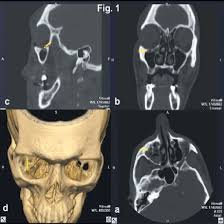
The images could be altered as a function of what the scanner achieves to offer an angled view of the area about an orbit. This is among the best ways to obtain a view of orbital fractures, tumors, infections, soft tissue abnormality, and whatever else by offering superior visualization. These 3D-type images would likely be assessed based on radiological analysis to accurately devise any kind of treatment.
This would allow assessment of surrounding areas, such as the sinuses and nasal cavity, as well as cranial nerves, in the context of a 3D CT Orbit scan for diagnosing certain conditions.
Indications for 3D CT Orbit
A 3D CT Orbit scan proves to be a diagnostic necessity that aids in diagnosing several orbital region pathologies affecting structures such as bones, muscles, nerves, and soft tissues around it. Below are the primary conditions that make a 3D CT Orbit scan a necessity:
- Orbital Trauma
Orbital Fractures This is the most common indication for 3D CT Orbit whenever there is trauma to the orbital region by accident, fall, or physical trauma, and it results in an orbital fracture. It shows good blowout fractures, the damage that results to the floor of the orbit or any fracture including bones and walls of the orbit.
Post-Traumatic Evaluation: With facial trauma, the 3D CT scan helps establish the severity of fractures and further tissue injury. These elements will then be used to plan and manage the injuries.
- Orbital Tumors and Masses
Benign and malignant tumours are also imaged with 3D CT Orbit with the help of a high-resolution 3D scanning device. Different benign conditions like lacrimal gland tumor, optic nerve glioma, or malignant conditions such as orbital lymphoma and metastasis are included.
Tumor Localization and Staging: It provides detailed information about the size, location, and spread of the tumor, which helps in planning the biopsy or surgical intervention and assessing the involvement of surrounding structures.
- Orbital Infections
Orbital Cellulitis is the inflammation and infection of tissues within the orbit. In such cases, 3D CT helps in identifying whether the infection has spread along with the presence of an abscess or other complications.
Sinus-related infections, including sinusitis may extend to the orbit. A 3D CT scan would take a view of the extent of the infection and how both the sinuses and tissues within the orbit are affected.
- Inflammatory and Neurological Conditions
Graves’ Orbitopathy, also referred to as Thyroid Eye Disease. Swelling and inflammation in extraocular muscles; they often perform an evaluation using 3D CT in determining the intensity of the enlargement of muscle with the quantity of fat available in the orbit.
Orbital Pseudotumor is an illness that takes the shape of inflammation of the orbit or Idiopathic Orbital Inflammatory Disease; it can be differentiated with the use of this 3D CT and possibly even guides in the treatment of the condition.
Orbital Neuropathies: If the suspected optic nerve compression or inflammatory conditions affect the nerve, then 3D CT imaging can identify structural abnormalities.
- Congenital and Developmental Abnormalities
Congenital Anomalies: A 3D CT scan is used in the identification of congenital anomalies of the orbit such as syndromes of the craniofacial and other structural anomalies that could predispose to orbital anomalies of development.
The scan is used in assessing the anomaly of orbital development. These include hypertelorism where the eyes are more abnormally wide-set, underdeveloped eyes often called microphthalmia, and so on.
Benefits of 3D CT Orbit
A 3D CT Orbit, or Three-Dimensional Computed Tomography of the Orbit, is better than the conventional methods of imaging and is a valuable tool in the assessment of the orbital region. Some of the advantages of this advanced imaging technique are:
- Comprehensive and Detailed Imaging
Detailed resolution high-view, 3D: An orbit CT scan with 3D CT Orbit will give unobstructed views of every point in the orbital region- three dimensions including the eye socket, orbital bones, extraocular muscles, optic nerve, the blood vessels, and surrounding soft tissues. This amount of detail would not be reachable by standard 2-D imaging; hence, one will be able to see such structures more clearly in the more complex area.
Multiple Angles: Due to the fact that the orbit could be seen from various angles and aspects, doctors can easily identify slight abnormalities, fractures, tumors, and other conditions not discovered in standard 2D X-rays.
- Improved Diagnostic Accuracy
Clearly outlines fractures and degree of fracture: 3D CT Orbit is good for blowout fractures, orbital fractures diagnosis, and display extent of injury. It may present with the involvement of several walls of the orbit as well as structures like sinuses getting involved, enhancing sensitivity.
It improves the detection of orbital tumors, which may be either benign or malignant. This imaging modality provides information concerning the size and location of the tumor and relationship of the tumor with the adjacent tissues. It is essential for achieving an accurate diagnosis and outlining treatment.
Soft Tissue Visualization: Contrast agents may sometimes enable visualization of soft tissues such as extraocular muscles, optic nerves, and vessels that might enhance the diagnostic sensitivity of conditions like orbital infections or inflammatory diseases.
- Surgical Planning and Precision
It gives the surgeon a more extended and precise anatomy of the orbit, so preoperative evaluation by 3D CT Orbit will guide the surgeon in planning the surgical procedure in fracture repair, tumor removal, or orbital reconstruction. This information will give him information about major structures, including the optic nerve and blood vessels, so they need to be preserved; therefore, risks from the surgical approach can be minimized.
Improved Surgical Results: Deep knowledge of orbital anatomy leads to more planned surgical act in the accomplishment of that, which in turn guarantees complete recovery with excellent results of the patients.
- Non-invasive and Fast
Non-Invasive: This 3D CT Orbit scan does not have incision or surgical procedures that help gather pictures regarding the orbits, so it will not cause the same extent of pain in patients getting diagnosed and decreases the risks since these are not considered invasive diagnostic tools.
It is fast and efficient; a whole scan usually takes only 10-15 minutes, hence a good, fast diagnostic tool in case emergencies call for time-sensitive information to decide the course of action.
- Accurate Monitoring and Follow-Up
Monitoring Disease Activity Since orbital tumors, orbital inflammation, and Graves’ orbitopathy are chronic diseases, the 3D CT Orbit can monitor over time the developing or changing nature of the diseases. Thus, a physician will be able to assess changes in tumor size, development of new lesions, or response to therapy.
Postoperative follow-up: Healing after surgery as of post-operative can be evaluated by 3D CT by ruling out complications like infection or recurrent tumor.