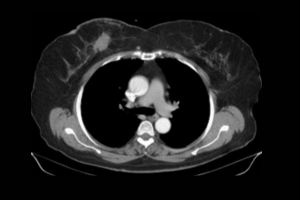
How CT Angiography of the Thorax Works
CT Angiography of the Thorax is one of the most advanced imaging procedures. CT, combined with the principles of angiography, takes accurate images of blood vessels within the chest. It scans images of the heart, the aorta, coronary arteries, and pulmonary arteries. The overview of the process is as follows:
You are asked to remove all your metal objects, including jewelry, glasses, or dentures, before starting the procedure since these can interfere with the scan.
You will be required to change into a hospital gown for the scan.
CT angiography employs the use of contrast dye, or contrast media, which is introduced into a vein in your arm via an intravenous line.
Contrast dye is used to highlight the blood vessels, making them white and prominent on the CT images. Thus, any abnormality in blood vessels can be easily picked up by the radiologist.
After administering the contrast, the patient is put on the CT scan table.
The table moves inside the CT scanner, a large, doughnut-shaped machine that revolves around the body to capture cross-sectional images.
The CT scanner takes multiple pictures with the help of X-rays from various angles. The computer combines all these images to provide 3D pictures of the thoracic blood vessels, thus allowing for a clear structure of their view.
- Respiratory Preparation :
You may be asked to hold your breath for short periods while you undergo the scan, as this helps reduce movement and allows images to be clearer when scanned. This is particularly critical when acquiring images of the heart and lungs; their integrity can be sensitive to movement.
The CT scanner captures a few quick X-ray images of the chest that are later reconstructed into high detail 2D and 3D images of the blood vessels in the thorax.
The images help a radiologist view the arteries and veins in detail so that they can easily determine whether there are blockages, aneurysms, pulmonary embolism, and other vascular disorders.
Indications for CT Angiography Thorax
Thorax CT Angiography is a very powerful diagnostic tool, checking the blood vessels in the chest, including the aorta, coronary arteries, pulmonary arteries, and other major vascular structures. Indications are mainly when any of the following conditions exist:
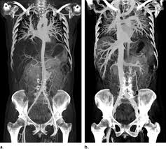
- Aortic Aneurysm and Dissection
Aortic Aneurysm: In the evaluation of the presence, size, and shape of an aneurysm within the aorta, particularly with suspicion of an enlarged or weakened artery.
Aortic Dissection: This is an observation and review of a tear in the inner lining of the aorta, a potentially lethal condition if not taken to attention.
- Coronary Artery Disease
Coronary Artery Stenosis: It detects the narrowing or blockages in the coronary arteries that carry blood to the heart. It can be used for the diagnosis of a patient who is experiencing chest pain, shortness of breath, and other symptoms that might be associated with heart issues.
Pre-Operative Evaluation: This will therefore help in determining the procedures that will be required, for example, CABG or even stenting, which require flawless morphological images of the vascular architecture of the coronary circulation.
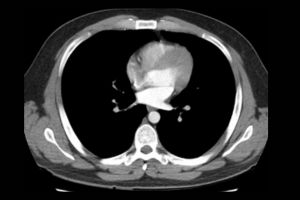
- Pulmonary Embolism (PE)
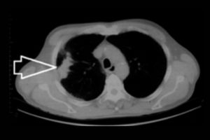
Pulmonary Embolism: CT Angiography is the primary scanning technique for pulmonary embolism, a potentially life-threatening illness due to a blood clot clogging up pulmonary arteries, thus hindering normal flow of blood in the lung.
It produces very brilliant images of pulmonary arteries and even detects even the smallest clots, which may be beyond the reach of other scanning methods
- Trauma or Injury
Vascular Injury: It identifies the occurrence of vascular injuries like hemorrhage or tears in the blood vessels of the chest following chest trauma, such as that of a car accident or a fall. It is mostly used in emergency areas for determining the severity of the injury.
Hemorrhage: It determines the bleeding inside the chest and into the surrounding blood vessels from the effects of blunt trauma.
- Congenital Vascular Abnormalities
Congenital Heart and Vascular Defects: It is useful in diagnosing and evaluating congenital vascular anomalies such as vascular malformations, aortic coarctation, and patent ductus arteriosus.
It is also useful in children as well as adults to visualize vascular anomalies that must be treated medically or surgically.
Benefits of CT Angiography Thorax
Thoracic CT Angiography has successfully proved to have offered several benefits that have made this radiologic procedure of value in the diagnosis and assessment of various vascular and cardiovascular disorders. These are as follows:
- Non-Invasive Procedure
No Surgery or Catheter InsertionProbably one of the best advantages of this specific type of radiological examination or technique called CT angiography is that it’s a purely non-invasive procedure. There is no insertion of catheters within the blood vessels, as in its previous counterpart labeled as traditional angiography does. This automatically means there is no possibility of infection or any chances of bleeding. No need for incision, subsequently reducing the scope of having complications in the process itself.
- Highly Detailed Ultra High-Resolution Images
Clear and detailed visualization: CT angiography provides clear, high-resolution images of the blood vessels in the chest with high resolution and clarity. This capability to create 3D allows the doctor to see the general image through the complex blood vessels, making it easier to identify anomalies in the blood vessels, including blockages, aneurysms, and vascular malformations.
The three-dimensional reconstructions give a good view of complex vascular anatomy that cannot be easily obtained with X-ray or ultrasound. These include detailed views of the aorta, coronary arteries, and pulmonary arteries.
- Quick and Easy
Rapid Process: The process of CT angiography is not that long. It takes 15 to 30 minutes, and the results are obtained within hours. So, it is a time-saving procedure as compared to other diagnostic procedures that would take a longer time or require more time.
Rapid Assessment: This is priceless in the immediate assessment of vascular structures. It is quite crucial in emergency cases such as pulmonary embolism or vascular injuries.
- Right Diagnosis
With CT angiography, doctors can accurately diagnose conditions like an aortic aneurysm, coronary artery disease, pulmonary embolism, and countless other vascular abnormalities. So, the doctor will be able to correctly direct treatment to be used either through surgery, medication, or changes in lifestyle.
Early Detection: The images are very detailed and can provide issues even before symptoms arise in most cases, thus allowing early intervention to occur in preventing what could have resulted in such life-threatening events as ruptures or strokes.
- Detailed Analysis
It can image most of the major arteries and veins within the chest area by starting from the aorta, moving along through the coronary arteries, pulmonary arteries, and pulmonary veins, thus giving a full view of the thoracic vascular system.
Multi-Condition Imaging in one Scan: It is very versatile where different conditions like that of coronary artery blockages, aortic aneurysms, pulmonary embolism can be tested in one sitting.
- Pre-Operative Planning
It also prepares for surgeries such as coronary artery bypass, stent placement, and aortic aneurysm repair. It gives clear images of the involved blood vessels. This way, surgeons prepare well for the surgery. This reduces the complications of the surgery and yields good results.
Monitoring post surgery: It also applied to monitor the outcome of surgeries and look out for complications which may arise in regard to restenosis, re-narrowing of the artery or graft failure.