
MRI Mammography and Spectroscopy
Book your MRI Mammography and Spectroscopy at Medifyhome, where we offer top-quality scans at the best prices. Our NABL & NABH accredited diagnostic centers ensure accurate and reliable results.


Book an Appointment
MRI Mammography and Spectroscopy
Medifyhome has collaborated with the best pathology laboratories that are NABL and NABH certified and follow ISO safety guidelines to provide the best MRI Mammography and Spectroscopy at an affordable price for needy individuals. Breast cancer continues to be a common type of cancer in females throughout the globe. In both cases early diagnosis plays a vital role in enhancing survival and prognosis. Mammography and ultrasound are common in the diagnosis of breast cancer, but they can be ineffective in revealing details required in women with dense breasts or those at high risk for developing breast cancer. This is where advanced methods of MRI Mammography, and MRI Spectroscopy pays their bills by providing more accurate and detailed methods than conventional mammography in the diagnosis of breast cancer.
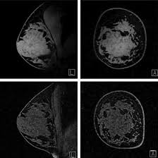
MRI Mammography (or Breast MRI) is a diagnostic study that utilizes a powerful magnetic field, radio waves, and digital signal processing to produce relatively high contrast pictures of the breast tissue. It is most useful for patients with high risk of developing breast cancer or patients with dense breast tissue, in which cases screen films may not detect small abnormalities. MRI Spectroscopy (MRS), on the contrary, performs beyond structural imaging. It evaluates the biochemical content of the kind of tissue, and from the change in the metabolism of this particular kind of tissue it may be able to identify the presence of cancer before the latter manifests structural deformities. It enables the early diagnosis of tumors and how breast cancer is likely to be in treatment.
To schedule an appointment for MRI Mammography and Spectroscopy, simply contact Medifyhome or call our customer care at +919100907036 or +919100907622 for more details and queries.
Overview
Specification
FAQ
Overview
MRI Mammography
MRI Mammography, or Breast MRI, is an advanced imaging technology used to create detailed pictures of the breast. This imaging technique differs from other mammograms in that they do not rely on X-rays but on powerful magnets and radio waves to make a high-resolution image of the breast. This creates a deeper understanding of breast health and can even detect abnormalities like tumors that might not be evident in other types of images.
How Does MRI Mammography Work?
It uses a magnetic field and radio waves to generate images of the inside of the breast. During the procedure:
- Contrast Injection: A special dye, known as contrast agent, is often injected into the bloodstream to help make the breast tissues more visible on the MRI scan.
- The process of imaging is done on an MRI table where a patient lies down face forward and places her breasts in a special coil. Hydrogen atoms are aligned in the body through the magnetic field, and the detailed images of the breast tissue are captured when the machine rotates.
- The results give the doctor the opportunity to know about the size, shape, and characteristics of any abnormality, hence giving them all the detailed structural information of the breast tissue.
Advantages of MRI Mammography
- Greater Sensitivity: Breast MRI excels in the identification of even the smallest tumor formations or abnormalities that are perhaps undetectable through any kind of traditional mammography, especially in cases involving a dense breast.
- It’s Non-invasive MRI Mammography is noninvasive and does not make use of ionizing radiation; that means it is much safer when repeated imaging is expected.
- Early Detection: It can detect breast cancer at an early stage, even when a lump or abnormality cannot be felt during a physical exam.
- Pre-surgical Planning: MRI Mammography is very helpful in assessing the spread of cancer before surgery so that doctors can decide the right treatment option.
When to have MRI Mammography?
Breast MRI is generally advised for:
- Women at Higher Risk: Family history of breast cancer, genetic mutation, and so on.
- Dense Breast Tissue: Women whose breasts are dense. It is often difficult to see the mammogram.
- Follow-up with Unclear Results from a Mammogram: A traditional mammogram is not clear. Then, an MRI can provide additional information.
- It monitors breast cancer. For patients diagnosed with breast cancer, MRI helps in monitoring how the cancer may have spread or returned.
MRI Spectroscopy
This imaging technique is known as MRS, or magnetic resonance spectroscopy. It provides additional detailed chemical information about the composition of tissues. In MRI spectroscopy, unlike with normal MRI, which simply is to look at anatomical structure, the metabolic and biochemical properties of the tissue under study are analyzed. Measurements of certain metabolites-such as choline, lactate, and creatine-can be determined. They can provide insight into a biochemical environment of tissue which in turn can identify cancers much before structural changes could take place.
How Does MRI Spectroscopy Work?
MRI Spectroscopy works with the same kind of magnetic fields and radio waves that are applied with conventional MRI but with more sophisticated sequences designed to collect information on metabolites.
- Magnetic Field and Radio Waves: Like with any other regular MRI, the patient lies down in a powerful magnetic field, where hydrogen atoms in the body become aligned along the direction of the magnetic field.
- Spectral Data Acquisition: Unlike creating a detailed image of the structure of the body, MRI Spectroscopy determines the concentration of different metabolites within the tissue by analyzing signals from hydrogen nuclei. The MRI scanner then produces a “spectrum” of such compounds.
- Metabolite Analysis: The spectrum shows the relative concentrations of various metabolites such as choline, creatine, lactate, and lipids. Some of these metabolites are elevated in cancerous tissue as well, though more specifically choline might be used as an indicator for the presence of tumor tissue since malignant tissue has different metabolic activity compared to normal tissue.
Benefits of MRI Spectroscopy
It could be employed for the early detection of cancer. MRS may be employed to detect the alterations of the metabolic condition of the tissue before the structural alterations possibly evident on the conventional MRI or mammography. It will, therefore, allow identifying the malignancies at an early stage.
- Non-invasive and ionizing radiation-free: MRS is non-invasive, as with MRI, and does not apply ionizing radiation, thus being safer for re-examination of sensitive patients.
- Distinguishing Benign from Malignant Lesions: MRS can differentiate between benign and malignant lesions through biochemical composition of the breast tissue and avoid unrequired biopsies and interventions.
- Determining Treatment Response: MRI Spectroscopy can be used to measure how tumors are responding to a treatment, providing real-time information regarding changes in metabolic activity of tissue.
- Applications of MRI Spectroscopy in Breast Cancer
- It may even detect biochemical changes that a malignant tumor may have without even showing any structural change within the conventional MRI. • Tumor Types: With MRS, invasive ductal carcinoma can be differentiated from other benign conditions like fibroadenomas or cysts.
When to Apply MRI Spectroscopy?
MRI Spectroscopy is applied when
- Unclear MRI or Mammogram Results: If the outcomes of a routine MRI or mammography are not clear, then MRS can provide further insight into the biochemical makeup of the tissue.
- Monitoring Known Tumors: In diagnosed breast cancer patients, MRS can be applied to the monitoring of the treatment effectiveness and possible recurrence.
- High-Risk Screening In women with dense breast tissue, or in the high risk of developing breast cancer, MRS can serve as an adjunct tool alongside other imaging techniques.
Comparison of MRI Mammography and Spectroscopy
MRI Mammography and MRI Spectroscopy are advanced imaging technologies applied in the diagnosis and monitoring of breast cancer but serve different purposes and provide different advantages. The differences, similarities, and complementarity of these technologies can be described as follows:
- Purpose and Focus
MRI Mammography:
- Focus: Primarily applied for imaging the structural anatomy of the breast.
- Purpose: It detects abnormalities such as tumors, cysts, or any other structural alterations in the breast tissue. It is particularly helpful for women who have dense breast tissue where traditional mammograms may not be very reliable.
MRI Spectroscopy:
- Focus: Focuses on the biochemical and metabolic composition of tissues rather than their structure.
- It describes changes in the breast tissue which can be initiated by the chemical alteration through for example quantification of some of the known metabolites such as lactate, choline and creatine. This makes it very easy to identify changes which may be metabolic changes in nature that could even herald structural changes.
- Technology and Mechanism
MRI Mammography
- This technology uses a super fortified magnetic field plus radio waves with a view to having better pictures of the architecture of the mammary gland.
- Usually makes use of a contrast agent which, for example, consists of dye that makes clearer the images of breast tissue.
- It provides detailed resolution images of the architecture of the breast, which gives one an indication of size, shape, and location of mass and lesions.
MRI Spectroscopy:
- This also employs magnetic fields and radio waves, but this time it is designed to detect chemical signals from the tissue rather than imaging its structure.
- Captures the metabolic profile of breast tissue based on the concentration of diverse metabolites, such as choline, lactate, and creatine, so that normal and abnormal tissues can be differentiated.
- Applications
MRI Mammography:
- Primary Application: Screening and Diagnosis of breast cancer, in at risk patients or in women with dense breasts.
- Secondary Application: Staging of known breast cancers, follow-up after treatment for recurrence.
MRI Spectroscopy:
- Primary Application: Assessment of metabolic changes within tissues, that could be an early indicator of cancer development or malignancy.
- Secondary Application: Tumor aggressiveness, benign or malignant lesion, and cancer treatment response.
- Sensitivity and Specificity
MRI Mammography:
- Very sensitive in the diagnosis of breast cancer, especially in patients with dense breasts where regular mammograms fail to detect the abnormalities.
- Sometimes it results in false positives, thus causing unnecessary biopsies or further investigations.
MRI Spectroscopy:
- Sensitive in the detection of metabolic changes suggestive of cancer even before the structural changes are evident.
- Helps identify the biochemical signature of malignancy, providing information on the nature and aggressiveness of the tumor.
- It can detect abnormalities very early, but the spectra are not easy to interpret. The false positives are less than in MRI Mammography.
Diagnostic Advantages
MRI Mammography:
- Best suited for showing structural changes like tumors, cysts and masses, and it is much better to represent women who have dense breast tissue than traditional mammography.
- Detection: May be helpful to identify small or inconspicuous lesions which would be missed by routine mammography.
MRI Spectroscopy:
Adds biochemical information that is helpful when the diagnosis with MRI Mammography is suggestive but not certain or in case more information is required about the nature of the lesion.
Assess the shift in metabolism due to treatment as a means of providing feed-back on the response of treatment.
Potentially be used in tumor detection even before MRI mammography, as this technique can sense the alterations in metabolism that do not necessarily have an observable structural change.
Specification
- Test Type: MRI Mammography and Spectroscopy
- Preparation:
- Wear a loose-fitting cloth
- You may need to fast for 4–8 hours before an MRI
- Carry Your ID Proof
- Prescription is mandatory for patients with a doctor’s sign, stamp, with DMC/HMC number; as per PC-PNDT Act
- Reports Time: With in 4-6 hours
- Test Price: Rs.8000
FAQ
How to book an appointment for an MRI Mammography and Spectroscopy?
To schedule an appointment for MRI Mammography and Spectroscopy, simply contact Medifyhome or call our customer care at +919100907036 or +919100907622 for more details and queries.
What is the difference between MRI Mammography and MRI Spectroscopy?
- MRI Mammography or Breast MRI is basically used to make detailed images of the structure of a breast using strong magnetic fields and radio waves. It mainly deals with the detection of tumors, cysts, etc., which are structural abnormalities.
- MRI Spectroscopy (MRS) examines the metabolic and biochemical properties of breast tissue. It identifies alterations in tissue composition, which can be indicative of cancerous conditions even before any structural changes are noticeable.
Why is MRI Mammography used for specific patients?
MRI Mammography is used for
- Women with dense breast tissue wherein the mammograms may not work well.
- High-risk patients who have a family history of breast cancer and/or genetic mutations, like BRCA1 and BRCA2.
- Monitoring of known breast cancer, because it gives detailed images regarding the size of the tumors, their spread, and the response to treatment.
How does MRI Spectroscopy detect cancer?
MRI Spectroscopy identifies metabolic changes in the breast tissue. Metabolic activities of the cancerous tissues are typically different from normal tissue. By detecting specific metabolite levels, like choline, lactate, and creatine, MRS identifies areas that could potentially indicate malignancy, even before there is any structural change on an MRI.
Is MRI Spectroscopy safe?
Yes, MRI Spectroscopy is safe for most patients. Like MRI Mammography, it does not have ionizing radiation, and thus safer for repeated images. However, there are some people with implanted devices, such as pacemakers or cochlear implants, who are pregnant, and need to get prior permission from the doctor before taking the test.
How long will an MRI Mammography or MRI Spectroscopy procedure take?
MRI Mammography typically requires 30 to 45 minutes depending on the detailed images needed.
MRI Spectroscopy is a little longer as it involves acquisition of structural and metabolic data. The process can be up to 45 minutes or an hour, depending on the scope of the exam.
Are there risks to MRI Mammography or MRI Spectroscopy?
- Both procedures are considered safe. Still, there are some possible risks in using contrast agents and claustrophobia:
- Contrast agents: Sometimes, a contrast dye is used to make pictures clearer. Rare side effects may include allergic reactions or kidney problems in sensitive patients.
- Claustrophobia: Since the MRI machine is narrow, some patients may feel anxious or claustrophobic. Be sure to inform the healthcare team if you suffer from anxiety or discomfort with tight spaces.
- Metallic implants: If you have implants like pacemakers, some metallic devices, or other metal-based medical devices, MRI procedures are not recommended without first discussing this with your doctor.
Does MRI Spectroscopy replace other breast cancer screenings like mammograms or ultrasounds?
No, MRI Spectroscopy is not a replacement for mammograms or ultrasounds. It actually supplements them. For instance, even though MRI Mammography can give the most structural images of the breast, MRI Spectroscopy provides other biochemical information that could help determine early signs of cancer or how effective a treatment is.
Why Choose Medifyhome for MRI Mammography and Spectroscopy?
Medifyhome is an online medical consultant, which offers home services not only in your city but also in all major cities of India, such as Hyderabad, Chennai, Mumbai, Kolkata and others. This makes it easy for us to work with diagnostic centers that boast of having the most accurate equipment. The customer service for booking the appointment of the services is available 24/7 and Medifyhome also comes with instructions. Medifyhome has not only the best diagnostic centres, but it offers them at very cheaper prices. If you have been tested, you can promptly schedule an appointment with a health care service through our list of skilled physicians. For appointments for MRI Mammography and Spectroscopy, you can chat with us through Medifyhome or call our customer care at +919100907036 or +919100907622 for more information or inquiries.