Book an Appointment
MRI For Right Knee With Cartigram
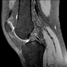
Medifyhome has collaborated with the best pathology laboratories that are NABL and NABH certified and follow ISO safety guidelines to provide the best MRI For Right Knee With Cartigram at an affordable price for needy individuals. The cartigram right knee MRI is the use of a technique through advanced imaging to highlight fine features in the area. Special MRI technique designed for showing much detailed images of the knee, in this case, especially looking at cartilage and all adjacent soft tissues. Cartigram with this form of an MRI advances and provides views which involve traditional MRI along with this cartigram special sequence. Cartilage is a very important element of the knee joint. It is essential for smooth movement. Any damage to this tissue, like tears or degeneration, leads to pain, stiffness, and restricted mobility. Whether it is a sports injury, arthritis, or other issues related to the knee, an MRI with Cartigram is a non-invasive means of assessing the health of your knee joint in great detail. Unlike X-rays or CT scans, MRI does not use radiation and provides high-resolution images of not only the cartilage but also ligaments, tendons, muscles, and bones, making it a powerful tool for diagnosing a variety of knee conditions.To schedule an appointment for a MRI For Right Knee With Cartigram, simply contact Medifyhome or call our customer care at +919100907036 or +919100907622 for more details and queries.
How Does MRI for Right Knee with Cartigram Work?
An MRI for the right knee with cartilage imaging, known as Cartigram, is a special kind of imaging. It is a combination of traditional Magnetic Resonance Imaging and advanced cartilage-sensitive imaging techniques used to assess the knee joint with particular attention to the cartilage and the surrounding structures. Here’s how it works:
- MRI Basics
MRI is an imaging procedure that uses a strong magnetic field and radiofrequency waves to produce detailed images of structures inside the body, including bones, muscles, ligaments, tendons, blood vessels, and soft tissues such as cartilage.
Unlike X-rays or CT scans, MRI does not utilize ionizing radiation, hence safer for multiple imaging and more appropriate for patients with chronic conditions or requiring frequent follow-up.
- Cartigram Technique
Cartigram is a dedicated imaging protocol or sequence under an MRI scan that emphasizes and evaluates the cartilage of joints, especially in the knee, articular cartilage.
The smooth, white tissue covers the ends of bones at a joint. This articular cartilage provides easy sliding movement with minimum friction. Damage to, degeneration of, or defects in cartilage can often be challenging to assess on routine MRI sequences, hence Cartigram.
- How the Cartigram MRI is Done
Preparation: The patient is usually asked to remove clothing with metal parts (like zippers) and will be put in a hospital gown. Any metal objects like jewelry, watches, or piercings have to be removed.
Positioning: You would lie on the MRI table. Your knee would be adjusted into a position that brings you and your knee nearer to the MRI machine with focus on the knee. You are usually positioned as for the knee to flex and rest in a slightly bent state.
MRI Scanning: The table will then slide into the MRI machine once the knee is in place. The MRI machine uses a strong magnetic field to create detailed cross-sectional images of your knee. The Cartigram-specific sequence is designed to enhance the visibility of cartilage, making any degeneration, thinning, or tears more visible. The scan may last between 20 to 40 minutes, depending on the complexity and area of the knee being studied.
- What Cartigram MRI Emphasizes
Cartigram imaging helps to throw out the following:
Cartilage Thickness: Whether the cartilage is thickened, normal, or thinned to assess degenerative conditions, such as osteoarthritis.
Cartilage Defects: The tears, lesions, or defects on the articular cartilage arising from injury or wear and tear.
Osteoarthritis and Joint Degeneration: Cartigram MRI is particularly useful for the early detection of osteoarthritis by revealing changes in cartilage integrity and signs of joint degeneration.
Chondromalacia Patellae: Cartigram can evaluate chondromalacia patellae, a condition wherein the cartilage under the patella or kneecap becomes damaged due to injury or overuse.
- Common Indications for MRI with Cartigram
This imaging technique is commonly used for:
Knee Pain: Recurring knee pain that is usually accompanied by inability to move the knee or stiffness of the joint.
Cartilage Injury or Damage: Suggested cartilage injury or damage, including tears, fractures, and lesions.
Osteoarthritis: Osteoarthritis progression, which is an illness where the cartilage gets worn out gradually.
Chondral Defects: Diagnosing chondral defects due to injuries or degenerative diseases.
What Conditions Can MRI for Right Knee with Cartigram Diagnose?
An MRI of the right knee with Cartigram is an advanced imaging technique applied primarily for the assessment of the articular cartilage of the knee joint as well as the surrounding structures. Here are the essential conditions that MRI of the right knee with Cartigram will be able to diagnose:
- Cartilage Damage and Degeneration
Osteoarthritis (OA):
MRI with Cartigram is very sensitive in demonstrating early osteoarthritis in the form of thinning or wear of cartilage, which characterizes the disease. Damages to the cartilage is one of the earliest features of OA and detailed visualization of integrity of cartilage through Cartigram helps in the staging of disease.
MRI may also demonstrate associated changes, such as bone marrow lesions or subchondral cysts, which are common in advanced osteoarthritis.
- Cartilage Tears or Defects
Chondral Defects:
Cartigram is very sensitive to chondral defects. Chondral defects include tears or lesions in the articular cartilage of the knee. Chondral defects may occur due to trauma, repeated stress, or degenerative process. Cartigram imaging demonstrates detailed images of cartilage tears, fissures, and irregularities, thus guiding physicians to determine the location and size of the defect.
Osteochondral Lesions:
- Knee Ligament and Meniscal Injuries (Indirect Evaluation)
While Cartigram is specifically designed to evaluate cartilage, MRI of the knee can also offer information about the ligaments and menisci in the knee. Although the majority of standard MRI sequences are used to evaluate the ligaments and menisci, Cartigram can indirectly assist in assessing the effect of cartilage injury on surrounding structures, including the ACL or medial and lateral menisci.
- Knee Joint Osteochondritis Dissecans (OCD)
Osteochondritis dissecans is a condition in which a piece of cartilage and the underlying bone in the joint becomes loose or detaches due to a lack of blood supply. MRI with Cartigram can identify OCD lesions, assessing the extent of the damage, and help guide treatment decisions.
- Knee Cartilage Regeneration and Post-Surgical Evaluation
Post-Surgical Monitoring:
It is also beneficial in the monitoring of cartilage repair after microfracture surgery, cartilage grafting, or stem cell injections using MRI with Cartigram. This allows assessment on whether cartilage is healing or regenerating adequately and if the surgical repair actually works in enhancing the growth of cartilage.
Benefits of MRI for Right Knee with Cartigram
An MRI for the right knee with Cartigram offers several significant benefits, especially for assessing cartilage and joint health in the knee. Here are the key benefits of this advanced MRI technique:
- Detailed Assessment of Cartilage Health
Visualization of Cartilage: The greatest strength of an MRI with Cartigram lies in its ability to depict very high-resolution images of articular cartilage of the knee. Cartigram facilitates enhancement of the visibility of cartilage, thereby facilitating improved detection of early damage to cartilage, thinning, or degeneration, which is less observable by a standard MRI.
- Non-Invasive and Radiation-Free
No Radiation: Since MRI does not use ionizing radiation like X-rays or CT scans, it is considered safer for patients who may require repeated imaging or those exposed to the cumulative effects of radiation.
Non-Invasive: MRI is completely non-invasive. There is no injection or surgical incision involved in the process, except perhaps when contrast might be used, which would make it a safer and more comfortable alternative compared to other invasive diagnostic procedures, such as arthroscopy.
- Detection of Problems in the Knee Joint Much Earlier
Early Cartilage Damage Detection: Cartigram MRI will identify early degeneration or damage to the cartilage before symptoms reach their full extent. Early detection would therefore help avert further damage in the joints. Patients who have been diagnosed early with osteoarthritis, chondromalacia patellae, or osteochondritis dissecans (OCD) are likely to experience a better outcome from these conditions through earlier intervention and treatment options like physical therapy, regenerative treatments, or minimal invasive surgical interventions.
- Advanced Detection of Cartilage Injury and Lesion
Cartigram MRI is also beneficial when done with athletes or any other victim experiencing knee injuries caused by accidents associated with sports or falling over.
It will show tears or contusions on the cartilage, thus influencing their joint functions.
A technique that detects osteochondral defects. Osteochondritis dise is found; it results from either chronic injury or trauma and can only involve destruction of both cartilage over bone.
- Cartilage Regeneration Assessment Post-Surgical Monitoring Cartigram MRI can best be used as a diagnostic tool to follow the regeneration of new cartilage after an intervention, which includes operations such as microfracture surgery, cartilage grafting, or even autologous chondrocyte implantation (ACI). Thus, it provides the knowledge of whether cartilage is regrowing and if so, healing well in that particular area.
Preparing for MRI for Right Knee with Cartigram
Preparing for an MRI on the right knee with Cartigram is relatively simple, but there are certain steps that must be taken in order to ensure a smooth and safe procedure.
- Clothing and Personal Items
Wear Comfortable Clothing: You will be required to change into a hospital gown as clothing containing metal parts, such as zippers, buttons, or underwire bras, could interfere with the MRI scan.
Remove All Metal Objects: The MRI machine uses strong magnetic fields, so it’s very important to remove all metal objects from your body, including:
Jewelry, Watches, hairpins, or hair clips, Eyeglasses, dentures and hearing aids
- Diet and Fluid Intake
Eat Normally (Usually): In most cases, you will not need to fast before an MRI for the knee. You can eat and drink as usual unless instructed otherwise.
Hydrate: Fluid restrictions are not strictly applied during a knee MRI, but staying hydrated will keep your body comfortable during the procedure.
Avoid Caffeine: If you are anxious or prone to claustrophobia, avoid excessive caffeine before the MRI, as it can make you more jittery or uncomfortable.
- Positioning and Comfort
Proper Positioning: When you get to the facility, the technician will give you instructions on how you should position your right knee. Normally, you would be lying on your back with the knee bent at a specific angle, but it would depend on the region you want imaged.
Pads and Supports: Should you need them, an MRI technologist would administer pads or supports to support your knee in order for it to be positioned most comfortably for imaging.
Remain as Still as Possible: Try to stay still throughout the MRI. This is crucial because any motions can create motion artifacts – blurry images. This requires some patience because the scan might take anywhere from 20 to 45 minutes, depending on the complexity and the number of sequences required.
- Process of the MRI Scan
The MRI Machine: Soon after you are comfortable, the table moves the patient into the actual machine. The MRI captures photographs of the knee joint and its respective cartilage using the strengths of a magnetic field and waves made from radio. A really loud knocking or thumping noise is common and always occurs during an MRI scanning procedure. You can even be provided with earplugs or headphones that decrease the noise.
- Test Type: MRI For Right Knee With Cartigram
- Preparation:
- Wear a loose-fitting cloth
- Fasting not required
- Carry Your ID Proof
- Prescription is mandatory for patients with a doctor’s sign, stamp, with DMC/HMC number; as per PC-PNDT Act
- Reports Time: With in 4-6 hours
- Test Price: Rs.5000
How can I book an appointment for a MRI For Right Knee With Cartigram through Medintu?
To schedule an appointment for a MRI For Right Knee With Cartigram, simply contact Medintu or call our customer care at +919100907036 or +919100907622 for more details and queries.
What is an MRI for Right Knee with Cartigram?
An MRI for the Right Knee with Cartigram is one of the specific types of MRI scans that focus on creating clear images of the knee joint, especially the cartilage. The “Cartigram” technique enhances the visibility of the cartilage, especially useful in the detection of tears, degeneration, or other damage that would not be visible on standard MRI scans. This method assists doctors in evaluating the cartilage’s health and, in turn, provides them with a better view of the conditions within the knee joint.
An MRI of the right knee with Cartigram is a safe procedure.
Yes, an MRI of the knee is very safe. It uses magnetic fields and radio waves to generate images, meaning no radiation is involved, unlike X-rays or CT scans. However, patients with certain implants, such as pacemakers, metal prosthetics, or clips, may not be eligible for an MRI. It’s essential to inform your healthcare provider about any metal implants or medical devices before the scan.
Does the MRI Procedure Hurt?
It hurts, the MRI itself; it doesn’t hurt to lay there for images to take. Images of your knee are going to be captured with the help of a rather large, loud MRI machine in a very large tube and during that time you should stay completely still. For some of the test there can be considerable discomfort if forced to be motionless, such as during the one taking your images.
How Long Does an MRI on the Right Knee Take?
Typically, it takes approximately 30 to 60 minutes for an MRI on the right knee. This can be even longer if you need several imaging sequences with or without contrast agents. The sequences that include Cartigram, for example, may add a few more minutes due to the additional images required for better cartilage visualization.
Do I Have to Undergo Contrast for My Right Knee MRI?
In some situations, a contrast agent might be administered to improve images of the knee joint. The contrast dye is injected intravenously into your arm. This may give clearer pictures of soft tissues such as cartilage, ligaments, and muscles. Your provider will tell you if the use of contrast is indicated for your specific MRI.
How should I prepare for my MRI?
Preparation for an MRI of the knee is usually straightforward. You may be asked to change into a gown and remove any metallic objects, such as jewelry, watches, or hairpins, before the scan. If a contrast agent is being used, you may need to avoid eating or drinking for a few hours before the procedure. Always follow the specific instructions provided by your healthcare provider.
Is MRI for the Right Knee with Cartigram Used for Diagnosing Cartilage Damage?
Yes, cartilage assessment by MRI can be very effective using this technique. The cartigram-enhanced imaging technique can visualize minor damage, degeneration, or tears of the knee cartilage, which sometimes cannot be visualized by basic MRI scans. It finds a lot of utility while diagnosing conditions such as chondromalacia patella, meniscus tears, and osteoarthritis.
Are There Any Risks with Contrast Dye in MRI?
Contrast agents used in MRI are generally safe, but some patients may experience mild side effects such as a metallic taste, warmth, or a slight headache. In rare cases, people may have an allergic reaction to the contrast dye, so it’s essential to inform your healthcare provider if you have a history of allergies to contrast agents or kidney problems.
Why Choose Medifyhome for MRI For Right Knee With Cartigram?
Medifyhome is an online medical consultant that provides home-based medical services not only in your area but also in most cities in India, including Hyderabad, Chennai, Mumbai, Kolkata, and more. We have collaborated with diagnostic centers that have the best machines and equipment to ensure you get accurate results. Medifyhome provides 24-hour customer service for booking the appointment of the services and guides you with instructions. Medifyhome also provides the best diagnostic centers at low prices. Once you receive your test results, you can easily book an appointment with our network of experienced doctors for consultation. To schedule an appointment for a MRI For Right Knee With Cartigram, simply contact Medifyhome or call our customer care at +919100907036 or +919100907622 for more details and queries.


