


Book an Appointment
MRI Upper Abdomen And MR Urography
Medifyhome has collaborated with the best pathology laboratories that are NABL and NABH certified and follow ISO safety guidelines to provide the best MRI Upper Abdomen And MR Urography at an affordable price for needy individuals. Magnetic Resonance Imaging (MRI) has become a vital tool in modern medicine, offering detailed images of internal structures without the use of ionizing radiation. Specifically, MRI of the upper abdomen provides clinicians with critical insights into various organs, including the liver, pancreas, kidneys, and gallbladder. This advanced imaging technique allows for the detection and diagnosis of conditions such as tumors, cysts, and inflammatory diseases, aiding in effective treatment planning. MR urography, a specialized application of MRI, focuses on the urinary tract, including the kidneys, ureters, and bladder. This non-invasive procedure is particularly useful in identifying issues such as kidney stones, obstructions, and anatomical abnormalities. By utilizing high-resolution imaging, MR urography enhances our understanding of urinary system pathologies, facilitating timely and accurate diagnosis. Together, MRI of the upper abdomen and MR urography represent a powerful combination for assessing a wide range of abdominal and urinary tract conditions, empowering healthcare providers to deliver targeted and effective care.
To schedule an appointment for an MRI Upper Abdomen And MR Urography, simply contact Medifyhome or call our customer care at +919100907036 or +919100907622 for more details and queries.
Overview
Specification
FAQ
Overview
What is an MRI?
The MRI is, therefore, a medical imaging technique that is used to make images of organs/tissues in the body. MRI is a technology that operates with strong magnets and radio waves in order to create images. According to this understanding, the MRT draws the hydrogen atoms in the body by dropping the magnetic field on them and through the radio waves interrupts this. And when the signal is switched off the hydrogen atoms release signals while getting back to normal position and these signals are used to build images. MRI uses high-resolution images that make it most suitable for capturing soft tissues, brain muscles and organs such as the thyroid gland. MRI is different from other techniques such as X-ray or computer tomography which utilise ionising radiation making MRI safer for many patients. MRI can be applied even to diagnose different disorders, such as tumours or flu, inflammation, and injuries. MRI is used to detect cancer, brain tumours, joint diseases, and spinal diseases. It can also be used to track the results of a disease or a treatment program.
MRI of the Upper Abdomen
MRI of the upper abdomen is a non-invasive imaging technique that provides high-resolution images of the abdominal organs, including the liver, pancreas, spleen, kidneys, and gallbladder. This advanced modality is particularly valuable in diagnosing various medical conditions, guiding treatment decisions, and monitoring disease progression.
Key Indications for MRI of the Upper Abdomen
MRI is typically recommended for:
1.Liver Disorders: Identifying tumors, cirrhosis, fatty liver disease, and other hepatic abnormalities.
2.Pancreatic Conditions: Evaluating pancreatitis, pancreatic tumors, and cysts.
3.Biliary Tract Issues: Assessing gallstones, bile duct obstructions, and cholangitis.
4.Renal Pathologies: Investigating kidney tumors, cysts, and other abnormalities.
5.Spleen Examination: Detecting splenomegaly or splenic lesions.
The Procedure for MRI of the upper Abdomen
- Preparation:
Patients may be asked to fast for several hours before the scan to ensure clear images.Inform the healthcare provider about any metal implants, allergies, or medical conditions.
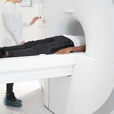
2. During the scan
Patients lie on a padded table that slides into the MRI machine. They may be given a contrast agent intravenously to enhance image quality. The scan typically lasts between 30 to 60 minutes
MR Urography
MR urography is a specialized imaging technique that uses magnetic resonance imaging to visualize the urinary tract, including the kidneys, ureters, and bladder. This non-invasive procedure provides detailed images, allowing for accurate diagnosis of various urinary system conditions.
Key Indications for MR Urography
- MR urography is commonly recommended for:
- Kidney Stones: Detecting the presence, size, and location of calculi.
- Urinary Tract Obstructions: Identifying blockages in the ureters or bladder.
- Anatomical Abnormalities: Evaluating congenital anomalies or changes in urinary tract structure.
- Tumors and Masses: Assessing renal or bladder tumors.
- Infections: Investigating complicated urinary tract infections or pyelonephritis.
The Procedure for MR Urography
- Preparation:
Patients may be advised to drink plenty of fluids before the exam to fill the bladder.
It’s essential to inform the healthcare provider of any allergies, particularly to gadolinium-based contrast agents, which may be used during the procedure.
2.During the scan
Patients lie on a padded table that moves into the MRI machine.The imaging process may take 30 to 60 minutes, during which patients need to remain still.In some cases, a contrast agent is injected intravenously to enhance the visibility of the urinary tract.
Interpreting Results
Radiologists analyze the images to identify any abnormalities, including:
- Presence of Stones: Size and location of any calculi.
- Obstructions: Areas where urine flow may be blocked.
- Tumors: Identification of any masses and their characteristics.
- Structural Issues: Anomalies in the anatomy of the kidneys or urinary tract.
Benefits of MRI and MR Urography
MRI and MR urography offer numerous advantages, making them essential tools in the diagnosis and management of various medical conditions. Here are some key benefits:
- Non-Invasive Imaging
Both MRI and MR urography are non-invasive procedures, allowing for detailed imaging without the need for surgical intervention.
- High-Resolution Images
MRI provides exceptional clarity and detail, particularly for soft tissues and organs, making it easier to identify abnormalities.
- No Ionizing Radiation
Unlike X-rays and CT scans, MRI does not use ionizing radiation, reducing the potential risks associated with radiation exposure, especially for repeated imaging.
- Comprehensive Evaluation
MRI of the upper abdomen allows for simultaneous assessment of multiple organs, providing a holistic view of abdominal health.
MR urography effectively visualizes the entire urinary tract, enabling the detection of various conditions in a single examination.
- Enhanced Contrast
The use of contrast agents in MRI and MR urography improves the visibility of structures and abnormalities, aiding in accurate diagnosis.
- Versatile Applications
MRI is useful for a wide range of conditions, including tumors, cysts, inflammatory diseases, and structural anomalies, while MR urography specifically targets urinary tract issues.
- Real-Time Imaging
MRI technology allows for the assessment of dynamic processes, such as blood flow, which can provide additional diagnostic information.
- Patient Comfort
MRI procedures are typically well-tolerated. Most patients experience minimal discomfort, and the process can be adjusted for individuals with anxiety or claustrophobia.
- Minimal Side Effects
The side effects of MRI and MR urography are generally minimal. Any reactions to contrast agents are rare and can often be managed effectively.
- Guiding Treatment Decisions
The detailed images obtained from MRI and MR urography play a crucial role in treatment planning, allowing healthcare providers to tailor interventions based on precise anatomical information.
Limitations and Considerations
While MRI and MR urography offer significant advantages, there are also limitations and considerations that patients and healthcare providers should be aware of:
- Contraindications
Metal Implants: Patients with certain implants, such as pacemakers, cochlear implants, or certain types of aneurysm clips, may not be suitable candidates for MRI due to the strong magnetic fields.
Pregnancy: Although MRI is generally considered safe during pregnancy, it is often avoided in the first trimester unless absolutely necessary.
- Claustrophobia and Anxiety
Some patients may feel anxious or claustrophobic in the MRI machine, which can affect their ability to remain still during the procedure. Alternative imaging options may need to be considered in such cases.
- Motion Artifacts
Any movement during the scan can lead to blurred images, potentially compromising the quality of the results. Patients must remain still, which can be challenging for some individuals.
- Duration of the Procedure
MRI scans can take longer than other imaging modalities, sometimes lasting 30 minutes to over an hour, which may be inconvenient for some patients.
- Cost and Availability
MRI and MR urography can be more expensive than other imaging methods, and access to MRI facilities may be limited in certain areas.
- Contrast Agent Reactions
While rare, some patients may experience allergic reactions to gadolinium-based contrast agents used during the procedure. It’s essential to discuss any known allergies or kidney issues with the healthcare provider beforehand.
- Limited Information for Certain Conditions
MRI is less effective in imaging structures with high calcium content, such as bones. For certain orthopedic conditions, CT scans may provide better information.
- Radiologist Expertise
The accuracy of MRI interpretation relies heavily on the expertise of the radiologist. Misinterpretation can lead to incorrect diagnoses or treatment plans.
- Availability of Advanced Techniques
Not all facilities may offer advanced MRI techniques (like functional MRI or MR angiography), which may limit the types of evaluations possible.
Advancements in MRI Technology
The field of magnetic resonance imaging (MRI) has seen significant advancements in recent years, enhancing its capabilities, efficiency, and overall effectiveness. Here are some key advancements in MRI technology:
- Higher Magnetic Field Strengths
Modern MRI machines often operate at higher magnetic field strengths (3.0 Tesla and above), providing improved image resolution and signal-to-noise ratios, allowing for more detailed imaging of soft tissues.
- Faster Imaging Techniques
Innovations like parallel imaging and compressed sensing have significantly reduced scan times, making procedures more efficient and comfortable for patients. These techniques minimize motion artifacts and enhance the overall workflow.
- Advanced Contrast Agents
Development of new gadolinium-based contrast agents and alternatives has improved the safety and efficacy of contrast-enhanced MRI. These agents can provide better visualization of certain tissues and abnormalities.
- Functional MRI (fMRI)
This advanced technique allows for the assessment of brain activity by measuring changes in blood flow. It is particularly valuable in neurology for mapping brain functions before surgical interventions.
- Diffusion Tensor Imaging (DTI)
DTI is used to visualize white matter tracts in the brain, providing insights into neurological conditions and injuries. This technique helps in understanding brain connectivity and assessing the impact of trauma or disease.
- Magnetic Resonance Spectroscopy (MRS)
MRS allows for the analysis of metabolic changes in tissues. It can provide valuable information about biochemical processes in tumors and other diseases, aiding in diagnosis and treatment planning.
- 3D Imaging Techniques
Advancements in 3D imaging allow for better visualization of complex anatomical structures. These techniques provide comprehensive views of organs and help in surgical planning.
- Artificial Intelligence and Machine Learning
The integration of AI and machine learning in MRI processing and analysis is revolutionizing the field. These technologies enhance image reconstruction, automate diagnostics, and improve interpretation accuracy by identifying patterns that may be missed by human observers.
- Portable MRI Systems
Development of portable MRI machines is making MRI more accessible, especially in emergency settings and remote locations. These systems can be used in critical care environments, improving patient care.
- Improved Patient Experience
Advances in magnet design and patient comfort features (e.g., wider bore MRI machines) are helping to reduce anxiety and improve the overall patient experience during scans.
Specification
- Test Type: MRI Upper Abdomen And MR Urography
- Preparation:
- Wear a loose-fitting cloth
- You will be asked to fast for 4 hours prior to your exam.
- Carry Your ID Proof
- Prescription is mandatory for patients with a doctor’s sign, stamp, with DMC/HMC number; as per PC-PNDT Act
- Reports Time: With in 4-6 hours
- Test Price: Rs.8000
FAQ
How can I book an appointment for an MRI Upper Abdomen And MR Urography through Medifyhome?
To schedule an appointment for an MRI Upper Abdomen And MR Urography, simply contact Medifyhome or call our customer care at +919100907036 or +919100907622 for more details and queries.
What is MRI, and how does it work?
MRI (Magnetic Resonance Imaging) uses strong magnetic fields and radio waves to create detailed images of internal organs and tissues. It works by aligning hydrogen atoms in the body and then measuring the signals emitted as they return to their original positions.
How long does an MRI scan take?
The duration of an MRI scan typically ranges from 30 minutes to an hour, depending on the complexity of the study and the specific areas being imaged.
Is MRI safe?
Yes, MRI is generally considered safe. It does not use ionizing radiation, which reduces the risk of radiation exposure. However, it’s important to inform your healthcare provider about any metal implants or other conditions that may affect your ability to undergo an MRI.
Do I need to prepare for the scan?
Preparation may vary. For MRI of the upper abdomen, you might be asked to fast for a few hours before the procedure. For MR urography, you may need to drink fluids beforehand to fill your bladder. Always follow the specific instructions provided by your healthcare provider.
Will I need a contrast agent?
In some cases, a contrast agent may be used to enhance the quality of the images. This is particularly common in MR urography. Inform your provider if you have allergies or a history of kidney problems, as this may influence the use of contrast.
What should I expect during the scan?
During the scan, you will lie on a padded table that slides into the MRI machine. You’ll need to remain still to obtain clear images. You may hear loud tapping or thumping noises during the scan, but headphones or earplugs are often provided to help minimize discomfort.
Can I have an MRI if I am pregnant?
While MRI is generally safe during pregnancy, it is often avoided in the first trimester unless absolutely necessary. Always discuss your situation with your healthcare provider.
What happens after the MRI?
After the scan, you can typically resume normal activities immediately. The images will be analyzed by a radiologist, who will provide a report to your healthcare provider, usually within a few days.
How are the results interpreted?
A radiologist will review the MRI images and provide a report detailing any findings. Your healthcare provider will discuss the results with you and recommend any necessary next steps.
What are the risks associated with MRI?
While MRI is safe for most people, there are some risks, including allergic reactions to contrast agents and the potential for discomfort from the enclosed space of the MRI machine. Always discuss any concerns with your healthcare provider before the procedure.
Why Choose Medifyhome for MRI Upper Abdomen And MR Urography?
Medifyhome is an online medical consultant that provides home-based medical services not only in your area but also in most cities in India, including Hyderabad, Chennai, Mumbai, Kolkata, and more. We have collaborated with diagnostic centers that have the best machines and equipment to ensure you get accurate results. Medifyhome provides 24-hour customer service for booking the appointment of the services and guides you with instructions. Medifyhome also provides the best diagnostic centers at low prices. Once you receive your test results, you can easily book an appointment with our network of experienced doctors for consultation. To schedule an appointment for an MRI Upper Abdomen And MR Urography, simply contact Medifyhome or call our customer care at +919100907036 or +919100907622 for more details and queries.