


Book an Appointment
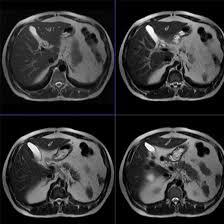
MRI Upper Abdomen With Contrast
Medifyhome has collaborated with the best pathology laboratories that are NABL and NABH certified and follow ISO safety guidelines to provide the best MRI Upper Abdomen With Contrast at an affordable price for needy individuals. Magnetic Resonance Imaging (MRI) of the upper abdomen with contrast is an advanced imaging technique that provides detailed views of the abdominal organs, including the liver, pancreas, kidneys, gallbladder, and spleen. This procedure enhances the visualization of soft tissues, allowing for more accurate diagnosis and assessment of various medical conditions. Using a gadolinium-based contrast agent, the MRI improves the differentiation between normal and abnormal tissues. This is particularly useful in identifying tumors, cysts, inflammation, and vascular abnormalities. The contrast agent highlights blood vessels and enhances the visibility of lesions, making it easier for healthcare providers to determine the nature and extent of any abnormalities.This non-invasive imaging technique is invaluable in various clinical scenarios, including the evaluation of liver diseases, pancreatic disorders, and urinary tract issues. Patients typically undergo this procedure after thorough preparation, which may include fasting and avoiding certain medications. By utilizing high-resolution imaging capabilities and advanced contrast techniques, MRI of the upper abdomen plays a crucial role in guiding treatment decisions and monitoring disease progression, ultimately contributing to improved patient care and outcomes.
To schedule an appointment for an MRI Upper Abdomen With Contrast, simply contact Medifyhome or call our customer care at +919100907036 or +919100907622 for more details and queries.
Overview
Specification
FAQ
Overview
What is an MRI?
The MRI is, therefore, a medical imaging technique that is used to make images of organs/tissues in the body. MRI is a technology that operates with strong magnets and radio waves in order to create images. According to this understanding, the MRT draws the hydrogen atoms in the body by dropping the magnetic field on them and through the radio waves interrupts this. And when the signal is switched off the hydrogen atoms release signals while getting back to normal position and these signals are used to build images. MRI uses high-resolution images that make it most suitable for capturing soft tissues, brain muscles and organs such as the thyroid gland. MRI is different from other techniques such as X-ray or computer tomography which utilise ionising radiation making MRI safer for many patients. MRI can be applied even to diagnose different disorders, such as tumours or flu, inflammation, and injuries. MRI is used to detect cancer, brain tumours, joint diseases, and spinal diseases. It can also be used to track the results of a disease or a treatment program.
What is Contrast in MRI?
MRI contrast agents are materials used to improve the visibility of particular structures and/ or fluids in the body during imaging. They assist radiologists get better and more enhanced images required in diagnosis of different ailments. Here we will be discussing the most commonly used contrast agents in the MRI. Gadolinium is an antigenic rare earth metal which can become safe for use in imaging, once it forms part of a chelated compound. However, wherever Gd is dominant, other types may be used in certain instances, such as iron oxide particles in imaging the liver. Contrast agents enhance the level of contrast reflected from normal and abnormal tissues. They make areas show increased blood flow or vascularity that is, tumours or lesions become more conspicuous. This is because with a boost in contrast images, the condition such as tumors infections and inflammatory diseases among others can easily be noticed. Which is introduced into veins – usually in the arm – directly to the bloodstream, where it spreads, and is taken up in tissues mainly by passive diffusion according to their vascularization and nature. MRI can sense the alterations in the signal intensity of the tissues that respond to the contrast, and yields better images.
Indications for MRI of the Upper Abdomen with Contrast
MRI of the upper abdomen in conjunction with contrast is a useful imaging technique employed routinely in the diagnosis of several pathologies affecting the liver, pancreas, biliary tree and some adjacent organs. Here are some common indications for using this imaging technique:
- Biliary Obstruction:
For diagnosing conditions that block the bile ducts, for example, the gallstones, tumor or stricture which may cause jaundice or cholangitis.
- Pancreatitis:
For initial evaluation or follow up of patients with an acute or chronic pancreatitis, to confirm or exclude complications, e.g. necrosis, pseudocyst or abscess formation.
- Tumors and Lesions:
For primary and metastatic tumors of the liver, the pancreas and in the biliary system including assessment of the tumor size, location, and the presence of vascular invasion.
- Liver Diseases:
As in cirrhosis, hepatitis, or fatty liver disease, this technique is useful to assess the consequences of the chronic liver disease on adjacent structures.
- Congenital Anomalies:
In patients with suspected congenital abnormalities of the biliary tree or pancreas that will impact function or are symptomatic.
- Vascular Assessments:
For visualisation of the hepatic artery and portal vein and other vascular structures to look for such pathologies as thrombosis or stenosis.
- Infection and Inflammation:
To visualize the abscesses or ongoing inflamed/inflammatory processes in liver, pancreas or the neighboring organs and tissues which are likely to need surgical management.
- Post-Surgical Evaluation:
In cases of biliary or pancreatic surgery or to diagnose leaks, strictures or abscesses in follow-up imaging.
- Assessment of Abdominal Pain:
MRI with contrast should be also used for the diagnosis of various upper abdominal pain when patients have no idea what causes it and other imaging methods do not reveal the problem.
- Guidance for Interventional Procedures:
For the purpose of offering precise presentations of the human anatomy that may be of help when performing certain procedures including biopsy or drainage.
Preparing for MRI with Contrast
Proper preparation for an MRI of the upper abdomen with contrast is essential to ensure a smooth and effective imaging experience. Here are the key steps to follow:
- Dietary Restrictions:
Fasting: You may be required to fast for 4-6 hours before the procedure. This helps ensure clear images and minimizes the risk of complications when using contrast.
Clear Liquids: In some cases, you may be allowed to drink clear fluids up to a few hours before your appointment. Always check with your healthcare provider for specific instructions.
- Medication Considerations:
Discuss Medications: Inform your healthcare provider about any medications you are taking, especially blood thinners, diuretics, or medications that affect kidney function.
Pre-Medication: If you have a history of allergic reactions to contrast agents, your provider may prescribe pre-medication to minimize the risk.
- Medical History:
Inform About Health Conditions: Share any medical conditions, particularly those related to kidney function, liver disease, or prior surgeries. This is crucial for assessing the safety of using contrast.
- Metal Objects and Implants:
Remove Metal Items: Before the MRI, remove all jewelry, watches, and other metal objects, as they can interfere with the magnetic field.
Inform About Implants: Notify the technician if you have any implanted devices, such as pacemakers, metal clips, or stents, as these may affect your eligibility for the procedure.
- Clothing:
Wear Comfortable Clothing: Dress in loose-fitting clothing without metal components. You may be asked to change into a hospital gown for the MRI.
- Addressing Anxiety and Claustrophobia:
Discuss Concerns: If you experience anxiety or claustrophobia, inform your healthcare provider. They may offer options for sedation or relaxation techniques to help you feel more comfortable during the procedure.
The MRI Procedure with Contrast
The MRI procedure with contrast for the upper abdomen is a comprehensive imaging process designed to provide detailed views of abdominal structures. Here’s what to expect during the procedure:
- Arrival and Check-In:
Arrive Early: Plan to arrive at the imaging facility 15-30 minutes before your scheduled appointment to complete any necessary paperwork.
Registration: You’ll check in at the front desk and may need to provide your insurance information and identification.
- Preparation:
Changing Clothes: You may be asked to change into a hospital gown to ensure that no metal objects interfere with the MRI.
Safety Screening: A technician will conduct a safety screening to check for any metal implants or other contraindications for the procedure.
- Positioning:
Lie Down on the MRI Table: Once in the imaging room, you’ll lie on a padded table. The technician will position you comfortably, typically on your back, and may use cushions for support.
- Administering the Contrast Agent:
IV Line Insertion: A healthcare professional will insert an intravenous (IV) line into a vein in your arm to administer the contrast agent.
Contrast Injection: The contrast agent, usually a gadolinium-based substance, will be injected through the IV. You may feel a brief sensation of warmth or coolness during this process.
- The MRI Scan:
Entering the MRI Machine: The examination table will slide into the MRI machine, which is a large, tube-shaped magnet. It may feel confined, but you will be able to communicate with the technician throughout the procedure.
Imaging Process: The MRI machine will begin taking images of your upper abdomen. You will hear loud thumping or tapping noises, which is normal. Earplugs or headphones may be provided to help reduce the noise.
Breath-Holding: During certain sequences, you may be asked to hold your breath for a few seconds to minimize motion and ensure clarity in the images.
- Duration of the Procedure:
Total Time: The entire MRI procedure typically lasts between 30 to 60 minutes, depending on the complexity of the imaging needed.
Interpreting MRI Results with Contrast
Interpreting MRI results with contrast involves analyzing the detailed images produced during the scan to diagnose various conditions affecting the upper abdomen. Here’s an overview of the process and what to expect:
- Role of the Radiologist:
Specialized Expertise: A radiologist, a physician trained in interpreting medical images, will review the MRI scans. They focus on identifying abnormalities in the liver, pancreas, biliary system, and surrounding structures.
Comparative Analysis: The radiologist may compare the current images with previous scans to assess any changes over time.
- Key Factors Analyzed:
Tissue Contrast: The use of contrast enhances the visibility of certain structures, allowing the radiologist to differentiate between healthy and abnormal tissues more effectively.
Lesion Characteristics: The size, shape, and signal intensity of any lesions or abnormalities are closely examined. This helps in determining if they are benign or malignant.
Vascular Structures: The radiologist assesses the blood vessels and ducts, checking for obstructions, abnormalities, or signs of inflammation.
- Common Findings:
Normal Results: If no abnormalities are found, the report will indicate that the structures appear normal, with no evidence of disease.
Abnormal Findings:
Biliary Obstruction: The presence of gallstones, strictures, or tumors causing blockages in the bile ducts.
Tumors: Identification of masses in the liver, pancreas, or biliary system, along with characterization of their nature (e.g., cystic vs. solid).
Pancreatitis: Signs of inflammation or complications, such as fluid collections or necrosis.
Cirrhosis: Changes in liver structure and texture indicating chronic liver disease.
- Reporting the Results:
Radiology Report: The radiologist compiles their findings into a formal report that includes a summary of observations, any diagnosed conditions, and recommendations for further evaluation or treatment.
Communication with Healthcare Provider: The report is sent to the referring physician, who will discuss the findings with you, explaining their implications and potential next steps.
- Follow-Up:
Depending on the findings, your healthcare provider may recommend additional tests, such as further imaging studies, biopsies, or consultations with specialists.
It’s important to schedule any follow-up appointments to discuss your results in detail and to understand any necessary treatments or interventions.
- Patient Understanding:
Discussing Results: Patients should feel empowered to ask questions about their MRI results, clarifying any medical terminology and understanding the implications for their health.
Understanding Limitations: While MRI with contrast provides valuable information, it may not capture every aspect of a condition, and further diagnostic procedures might be needed.
Benefits of MRI with Contrast
MRI with contrast offers several advantages, making it a valuable diagnostic tool for evaluating conditions in the upper abdomen. Here are the key benefits:
- Enhanced Image Quality:
Improved Visualization: The use of contrast agents significantly enhances the visibility of internal structures, allowing for clearer and more detailed images of soft tissues, blood vessels, and organs.
Differentiation of Tissues: Contrast helps differentiate between normal and abnormal tissues, making it easier to identify tumors, lesions, and other pathologies.
- Accurate Diagnosis:
Early Detection: MRI with contrast can detect abnormalities at earlier stages, which is crucial for effective treatment and better patient outcomes.
Characterization of Lesions: The ability to assess the characteristics of lesions—such as their size, shape, and vascularity—helps in determining whether they are benign or malignant.
- Non-Invasive Procedure:
No Ionizing Radiation: Unlike CT scans, MRI uses magnetic fields and radio waves, avoiding exposure to ionizing radiation, making it safer for patients, especially for those requiring multiple scans.
- Comprehensive Assessment:
Multi-Organ Evaluation: MRI with contrast provides a comprehensive view of the liver, pancreas, gallbladder, and surrounding structures, facilitating the evaluation of conditions affecting multiple organs.
Detailed Vascular Imaging: The technique can visualize blood vessels and assess conditions like vascular obstructions, which may not be as clearly seen on other imaging modalities.
- Guidance for Treatment Planning:
Informed Decision-Making: The detailed information obtained can guide healthcare providers in planning treatments, such as surgery, targeted therapies, or further diagnostic procedures.
Monitoring Treatment Response: MRI with contrast can be used to monitor the effectiveness of ongoing treatments, allowing for adjustments based on imaging findings.
- Safety and Tolerability:
Well-Tolerated: Most patients tolerate the procedure well, with minimal side effects from the contrast agents. Gadolinium-based contrasts are generally considered safe, although pre-screening for allergies and kidney function is essential.
- Versatile Applications:
Broad Range of Indications: MRI with contrast is used for various clinical indications, including assessing liver disease, pancreatic conditions, biliary obstructions, and abdominal pain of unknown origin.
Specification
- Test Type: MRI Upper Abdomen With Contrast
- Preparation:
- Wear a loose-fitting cloth
- Fasting required
- Carry Your ID Proof
- Prescription is mandatory for patients with a doctor’s sign, stamp, with DMC/HMC number; as per PC-PNDT Act
- Reports Time: With in 4-6 hours
- Test Price: Rs.8500
FAQ
How can I book an appointment for an MRI Upper Abdomen With Contrast through Medifyhome?
To schedule an appointment for an MRI Upper Abdomen With Contrast, simply contact Medifyhome or call our customer care at +919100907036 or +919100907622 for more details and queries.
What is an MRI with contrast?
An MRI with contrast is a specialized imaging technique that uses a contrast agent, typically a gadolinium-based substance, to enhance the visibility of internal structures during an MRI scan.
Why is contrast used in MRI?
Contrast agents improve image quality by highlighting specific tissues, blood vessels, and abnormalities, allowing for more accurate diagnoses of conditions in the upper abdomen.
Is the contrast agent safe?
Gadolinium-based contrast agents are generally safe for most patients. However, there is a risk of allergic reactions or complications in individuals with severe kidney impairment, which is why kidney function is typically assessed prior to administration.
What should I do to prepare for an MRI with contrast?
You may need to fast for 4-6 hours before the procedure. Discuss any medications you are taking and inform your healthcare provider about any allergies or kidney issues.
What will happen during the procedure?
You will lie on a padded table, and an intravenous (IV) line will be inserted to administer the contrast agent. The MRI machine will then take images of your abdomen while you may be asked to hold your breath during certain sequences.
How long does the procedure take?
The entire process usually lasts between 30 to 60 minutes, depending on the complexity of the imaging required.
Will the MRI with contrast hurt?
The procedure is generally painless. Some patients may feel a brief sensation of warmth or coolness during the contrast injection, but this is usually mild and temporary.
What are the risks associated with MRI with contrast?
While MRI is considered safe, potential risks include allergic reactions to the contrast agent and nephrogenic systemic fibrosis (NSF) in patients with severe kidney dysfunction. Always discuss any concerns with your healthcare provider.
How will I receive the results?
A radiologist will analyse the MRI images and generate a report. Your referring physician will discuss the findings with you and recommend any necessary next steps.
Can I eat or drink after the MRI?
Yes, you can typically resume your normal activities, including eating and drinking, immediately after the procedure. Drinking fluids is encouraged to help flush the contrast agent from your system.
Why Choose Medifyhome for MRI Upper Abdomen With Contrast?
Medifyhome is an online medical consultant that provides home-based medical services not only in your area but also in most cities in India, including Hyderabad, Chennai, Mumbai, Kolkata, and more. We have collaborated with diagnostic centers that have the best machines and equipment to ensure you get accurate results. Medifyhome provides 24-hour customer service for booking the appointment of the services and guides you with instructions. Medifyhome also provides the best diagnostic centers at low prices. Once you receive your test results, you can easily book an appointment with our network of experienced doctors for consultation. To schedule an appointment for an MRI Upper Abdomen With Contrast, simply contact Medifyhome or call our customer care at +919100907036 or +919100907622 for more details and queries.